Recrystallization and grain growth in minerals: recent developments
Janos L. Urai
1 and Mark Jessell21
Geologie- Endogene Dynamik, Fachgruppe Geowissenschaften, Lochnerstrasse 4-20, D-52056 Aachen, Germany.2
Department of Earth Sciences, Monash University, Box 28E, Melbourne, Victoria, 3800, Australia;Keywords: dynamic recrystallization, grain growth, minerals, geoscience.
Abstract. Recent developments in the study of recrystallization and grain growth in rocks and minerals are briefly reviewed and key issues discussed.
1 Introduction
The study of deformation mechanisms and microstructure evolution in geosciences is different from that in engineering [1]. Firstly, we aim to reconstruct the processes and conditions in the geologic past from observations on rocks which are brought to the Earth's surface by geologic processes or by deep drilling [2, 3] or to infer such processes operating at present in the deep subsurface using measurements of acoustic or electric response of the lithosphere [4]. The second aim is to provide constitutive models which can be used as input for the modelling of geodynamic evolution [5-7].
The very low strain rates in geologic deformation (10-13 s-1 is a common value) mean that these natural processes are not directly accessible by experiment. Therefore Earth materials scientists combine extrapolations of laboratory data [8-14] with observations of both naturally and experimentally deformed rocks and theoretical modelling [15-19].
In the sixties these studies were strongly influenced by the metallurgical and materials science literature [20-22]. In the past four decades understanding of the subjects has increased dramatically, and the limits of applicability of concepts from engineering materials were clarified. With the increase of data available and the improvement of analytical tools, communication between experimentalists and structural geologists has become increasingly challenging. Laboratory studies focus more on quantifying the behaviour of simple, pure or synthetic rocks under well defined conditions [23-27]; studies addressing the behaviour of polyphase rocks [28-30] are less frequent. On the other hand, studies of naturally deformed and recrystallized materials [31-37] emphasize the complexities of the processes occurring in nature which often involve changes in chemical composition and complex deformation paths under changing temperature and pressure.
The aim of this paper is to give a brief overview of the developments and issues in the study of recrystallization and grain growth in minerals over the past fifteen years, using a selection of papers from the large body of literature in this rapidly evolving field.
2 Dynamic recrystallization
Displacements of the Earth's crust and upper mantle are accommodated at least in part by shear zones in which very large strains are accumulated in zones of localized noncoaxial deformation [38, 39]. During localization of strain in polyphase rocks softening mechanisms involve the rearrangement of phases by plastic flow and dynamic recrystallization, producing a change of the load-supporting phase [28, 40] or in some cases a change in deformation mechanism to grain size sensitive flow by a grain-scale mixing of the two phases [41].
Dynamic recrystallization is a common process in ductile shear zones [42-45], in fact the vast majority of recrystallization processes in the Earth are dynamic in nature.
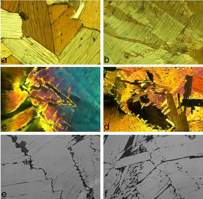
The microscale mechanisms of dynamic recrystallization are rotation of subgrains until new high angle grain boundaries are formed [46, 47], and grain boundary migration [48, 49]. Fig. 1 is an illustration of dynamic recrystallization in biotite, with kinking due to the presence of only one easy slip system and grain boundary migration in the presence of a small amount of partial melt in the grain boundaries.

Fig. 1. Experimentally deformed biotite, a highly anisotropic sheet silicate . Samples were deformed at 1 GPa pressure with about 3% H2O-rich fluid added. (a) Starting material, a coarse grained natural biotite rock. Transmitted light micrograph, horizontal edge of photo is 5 mm. (b) Deformed grain showing kinks developed to accommodate deformation by basal glide which is the only easy slip system in Biotite. Black lines are microfractures along the basal plane. Transmitted light micrograph, horizontal edge of photo is 1 mm. (c) Kinks in a grain deformed at 10-6 s-1 at 800 C, for 90 hours. Incipient recrystallization by kink boundary migration. Transmitted light micrograph, horizontal edge of photo is 1 mm. (d) Dynamically recrystallized grains in a sample deformed 20,000 hours at 10-7 s-1 at 820 C. Anisotropic growth of new grains occurs along former grain boundaries. Transmitted light micrograph, horizontal edge of photo is 1 mm. (e, f) SEM BSE micrographs of the sample shown in (d), showing kink boundary migration and growth of new grains which have a significantly different composition from the parent grains. Dark grey areas are the small amounts of melt which formed along preexisting grain boundaries and facilitated grain boundary migration. Horizontal edge of photos is 0.1 mm.
As a result of dynamic recrystallization, a transition to grain size sensitive flow may occur, during phases of transiently low grainsize after a previous period of dynamic recrystallization at high shear stress [50] or caused by stabilization of the grainsize by second phases [51]. The conditions under which the changes in grain size during dynamic recrystallization can lead to a switch in deformation mechanism to grain size sensitive creep and consequent localization of strain has been discussed extensively in the Earth Science literature. Recently, an interesting new model was presented by de Bresser and coworkers [52, 53]. According to this model, dynamic grain growth and grain size reduction during dynamic recrystallization lead to a balance at steady state such that the strain rate contributions of dislocation and diffusion creep become equal. This means that during steady state dynamic recrystallization there will be a significant contribution of diffusionally accommodated grain boundary sliding. It also means that dynamic recrystallization in a pure material cannot produce a switch in deformation mechanism to dominantly grainsize sensitive creep. During geological deformation under changing temperature and strain rate however, it is possible that steady state is not reached and large part of the rock's history are dominated by transient effects. Research into these transient effects is still in its infancy.
3 Transmitted light deformation of model materials
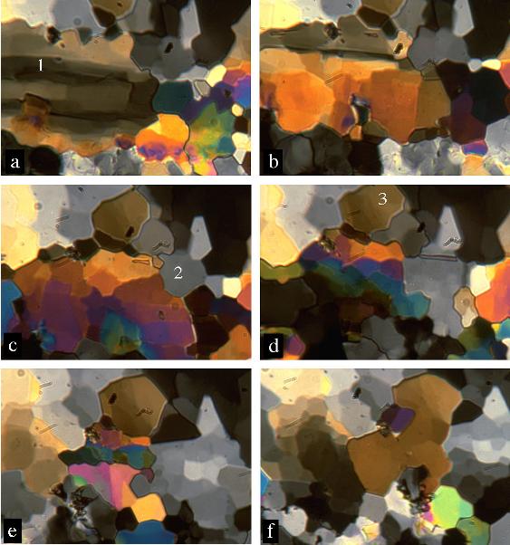
Transparent, optically anisotropic crystalline materials with a low melting point can be deformed in small deformation cells in plane strain under the polarized light microscope [11]. These experiments allow measurement of the full grain scale displacement field [55], together with real time observation of the evolving microstructure and the change in orientation of the crystal lattice at every point in the sample [56, 57]. Time lapse movies made from such experiments [58] allow detailed study of grain scale microstructure evolution [59, 60] and measurement of grain boundary velocity [61] [62]. In some cases the experiments are scaled models of much higher temperature deformation of minerals because the model material belongs to the same isomechanical group [62]. An example of a transmitted light deformation experiment is shown in Fig. 2.
With the rapid development in the numerical modelling of microstructural evolution [63] transmitted light deformation experiments are becoming an important tool for the validation of such models. Using the properties of the model material and the boundary conditions of the experiment, the digitized microstructure of the starting material can be used as input for simulations. Results of the simulations can be compared with measurements of displacement, lattice orientations and microstructure in the experiments.

Fig. 2. Sequence of images from a transmitted light deformation experiment of polycrystalline NaNO3 at 0.98 of its melting temperature . The images were taken during simple shear deformation at a strain rate of 10-5 s-1, over a period of about one hour. The microstructural evolution consists of the rapid growth of large new grains (1, 2 and 3) nucleated from pre-existing small grains, followed by rotation recrystallization by progressive misorientation of subgrains, and slow migration of these new high angle boundaries. Crossed polarizers, width of each image is 0.5 mm.
4 Grain boundary structure
Changes in grain boundary structure in rocks have large effect on mechanical and transport processes [64, 65] and fast grain boundary diffusion coupled with grain boundary migration is an effective process to change the chemical composition of grains, with the difference in composition between old and new grains providing a possible additional driving force for grain boundary migration [66].Our understanding of the structure and mobility of grain boundaries in minerals is poorly constrained. This partly due to the experimental difficulties and also to the large variability in grain boundary structure in recrystallizing rocks, from clean, solid-state boundaries [67] to micron-wide zones of fluid or melt between two grains. Such fluid-filled grain boundaries migrate by dissolution of the grain with the higher free energy, diffussion across the fluid and crystallization on the other side [21]. The morphology of the fluid in these grain boundary films has been proposed to be different from the morphology in static equilibrium [68-73], but more research is needed for a satisfactory resolution of this issue.
5 Grain growth
Because of the low driving forces during static grain growth, very small amounts of deformation can have a significant effect on the evolving microstructures. Grain growth in nature is frequently accompanied by deformation [74, 75] and the perfect foam textures which form during static grain growth of metals are infrequent in rocks. Examples of close-to static settings are the contact aureoles of granite intrusions [76].
Laboratory studies of grain growth [27, 77, 78] are in part also directed to investigating dynamic increase in grainsize [79].
The well known effects of an insoluble second phase on the pinning of grain boundaries and stabilizing grainsize are also common in rocks [24, 51, 80-82] although quantitative understanding of their effect needs much further study.
6 The relationship between recrystallization, mechanical properties and CPO
The highly anisotropic crystal structure of most minerals results in anisotropic bulk properties of textured materials undergoing dislocation creep. To understand the flow of such rock masses we need to quantify the anisotropic constitutive law of the material as a function of temperature, impurity concentration and deformation history, and implement this in a numerical model to simulate the deformation in nature. Sufficient data to attempt this are rarely available for rocks. An example is the flow of ice in polar ice sheets. Here an unusually complete dataset is available consisting of well characterized cores with a well known history of simple coaxial deformation over 400,000 years. Measurement of the grainsize and dislocation density along the cores shows good correspondence with modelling [83-85]
During burial to several hundred metres depth the ice undergoes grain growth during slow deformation, driven by the reduction in grain boundary energy. The rate of grain growth correlates with small variations in impurity levels caused by climate cycles in the past [86]. At further burial and deformation the ice undergoes rotation recrystallization which prevents further increase of the grainsize [87]. A strong CPO is developed, with an associated increase in mechanical anisotropy which in turn influences the displacement field in the ice sheet.
In the bottom 100 m of the ice sheet temperatures are sufficiently high (above -10 C) to cause a dramatic increase of grain boundary mobility and a three orders of magnitude increase in grain boundary migration rate [88]. In this regime grain boundary migration recrystallization is the dominant process, producing large lobate grains and a reduction of the strength of the CPO.
7 Interpretation of structures in naturally recrystallized rocks
Building on the rapidly growing body of knowledge of which this paper gives a brief overview, modern studies of naturally deformed rocks are capable of reconstructing considerable detail of the thermal and deformational history using detailed microstructural and mineralogical study [89-94].
On the other hand, some of these interpretations are not accepted by all, as quite different microscale processes can produce strikingly similar microstructures [95], and the complex overprint on a microstructure during a rock's journey to the surface is not well understood.
One of the methods to reconstruct the stress state in the past from microstructural information is based on the well known relationship between dynamically recrystallized grainsize and shear stress. Based on laboratory calibrations of this relationship for minerals, paleopiezometers are now in use for several minerals [53, 96, 97]. Recent work has shown that, in contrast to previous models, the temperature dependence of the grainsize-stress relationships cannot be neglected, and for improved paleostress estimates more extensive characterization of the deformation conditions is required.
An important development in analytical techniques is the possibility to create orientation images, using EBSD [98, 99] or optical techniques [100, 101]. This allows rapid access to the full richness of microstructural information in a rock, and has the promise to bring new diagnostic microstructures to improve our interpretation capabilities. The first studies in this field use boundary hirarchy characteristics to distinguish the dominant deformation mechanisms [102-104].
References
[1] S. M. Schmid, R. Heilbronner, H. Stünitz, Tectonophysics 303, vii-ix (1999).
[2] R. J. Knipe, Journal of Structural Geology 11, 127-146 (1989).
[3] P. J. O'Brien, J. Duyster, B. Grauert, B. Stöckhert, K. Weber, Journal of Geophysical Research 102 B8, 18203-18220 (1998).
[4] S. Zhang, S.-i. Karato, J. Fitz Gerald, U. H. Faul, Y. Zhou, Tectonophysics 316, 133-152 (2000).
[5] R. N. Pysklywec, C. Beaumont, P. Fullsack, Geology 28, 655-658 (2000).
[6] S. Ellis, S. Wissing, A. Pfiffner, Int. J. Earth Sci. in press (2001).
[7] M. R. Handy et al., Int. J. Earth Sci. in press (2001).
[8] M. S. Paterson, Tectonophysics 133, 33-43 (1987).
[9] G. Hirth, J. Tullis, Journal of Structural Geology 14, 145-159 (1992).
[10] L. N. Dell'Angelo, J. Tullis, Tectonophysics 169, 1-21 (1989).
[11] W. D. Means, Journal of Structural Geology 11, 163-174 (1989).
[12] M. S. Paterson, International Journal of Earth Sciences in press (2001).
[13] G. Hirth, C. Teyssier, W. J. Dunlap, Int. J. Earth Sci in press (2001).
[14] W. Skrotzki, R. Tamm, C.-G. Oertel, J. Röseberg, H.-G. Brokmeier, Journal of Structural Geology 22, 1621-1632 (2000).
[15] J. P. Busch, B. A. van der Pluijm, J. Struct. Geol. 17, 677-688 (1995).
[16] C. Pauli, S. M. Schmid, R. P. Heilbronner, Journal of Structural Geology 18, 1183-1203 (1996).
[17] R. L. M. Vissers, M. R. Drury, J. Newman, T. F. Fliervoet, Tectonophysics 279, 303-325 (1997).
[18] H. R. Wenk, G. Canova, Y. Bréchet, L. Flandin, Acta Materialia 45, 3283-3296 (1997).
[19] D. L. Olgaard, Geol. Soc. Spec. Pub. 54 (1990) pp. 175-181.
[20] Poirier, Creep of Crystals (Cambridge University Press, 1985).
[21] J. L. Urai, W. D. Means, G. S. Lister. (1986) .AGU Geophysical Monograph 36, pp. 161-199.
[22] M. R. Drury, J. L. Urai, Tectonophysics 172, 235-253 (1990).
[23] S.-I. Karato, Phys. Earth Planet. Interiors 51, 107-122 (1988).
[24] D. L. Olgaard, B. Evans, Contrib. Min. Petrol. 100, 246-260 (1988).
[25] G. C. Gleason, J. Tullis J. Struct. Geol. 15, 1145-1168 (1993).
[26] S. J. Covey-Crump, Journal of Structural Geology 19, 225-241 (1997).
[27] G. Dresen, Z. Wang, Q. Bai, Tectonophysics 258, 251-262 (1996).
[28] L. Dell'Angelo, J. Tullis, Tectonophysics 256, 57-82 (1996).
[29] J. Tullis, H. Stünitz, C. Teyssier, R. Heilbronner. (2000), http://www.virtualexplorer.com.au/VEjournal/Volume2.
[30] P. D. Bons, J. L. Urai, Tectonophysics 256, 145-164 (1996).
[31] R. J. Cumbest, M. R. Drury, H. L. M. v. Roermund, C. Simpson, Contributions to Mineralogy and Petrology 101, 339-349 (1989).
[32] R. Kruse, H. Stünitz, Tectonophysics 303, 223-249 (1999).
[33] T. F. Fliervoet, S. H. White, J. Struct. Geol. 17, 1095-1109 (1995).
[34] D. J. Prior, J. Wheeler, Tectonophysics 303, 29-49 (1999).
[35] J. C. White, C. K. Mawer, J. Struct. Geol. 8, 133-143 (1986).
[36] J. Newman, W. M. Lamb, M. R. Drury, R. L. M. Vissers, Tectonophysics 303, 193-222 (1999).
[37] U. Altenberger, S. Wilhelm, Tectonophysics 320, 107-121 (2000).
[38] R. L. M. Vissers, M. R. Drury, Tectonophysics 249, 155-171 (1995).
[39] E. H. Rutter, Tectonophysics 303, 147-158 (1999).
[40] K. Schulmann, B. Molcoch, J. Struct. Geol. 18, 719-733 (1996).
[41] J. H. Behrmann, D. Mainprice, Tectonophysics 140, 297-305 (1987).
[42] D. Jin, S.-I. Karato, M. Obata, J. Struct. Geol. 20, 195-209 (1998).
[43] G. Molli, P. Conti, G. Giorgetti, M. Meccheri, N. Oesterling, Journal of Structural Geology 22, 1809-1825 (2000).
[44] M. Bestmann, K. Kunze J. Struct. Geol. 22, 1789-1807 (2000).
[45] N. S. Mancktelow, Tectonophysics 135, 133-153 (1987).
[46] O. Nishikawa, T. Takeshita, J. Struct. Geol. 22, 259-276 (2000).
[47] F. Heidelbach, K. Kunze, H.-R. Wenk, J. Struct. Geol. 22, 91-104 (1999).
[48] B. Lafrance, B. E. John, B. R. Frost, J. Struct. Geol. 20, 945-955 (1998).
[49] T. Takeshita, I. Hara, Journal of Structural Geology 20, 417-431 (1998).
[50] L. Dell'Angelo, D. Olgaard J. Geophys. Res. 100, 15425-15440 (1995).
[51] M. Krabbendam, J. L. Urai, J. of Structural Geology in press (2001).
[52] J. H. P. de Bresser, C. J. Peach, J. P. J. Reijs, C. J. Spiers, Geophysical Research Letters 25, 3457-3460 (1998).
[53] J. H. P. de Bresser, J. H. ter Heege, C. J. Spiers, International Journal of Earth Sciences in press (2001).
[54] J. L. Urai, R. L. M. Vissers, Abstract, Leeds Conf 1988. (1989).
[55] P. D. Bons, M. W. Jessell, J. of Structural Geology 21, 323-334 (1999).
[56] M. Herwegh, M. R. Handy, R. Heilbronner, Tectonophysics 280, 83-106 (1997).
[57] R. Heilbronner, M. Herwegh, J. of Struct. Geol. 19, 861-874 (1997).
[58] J. L. Urai, F. J. Humphreys. (2000), www.virtualexplorer.com.au/ VEjournal/Volume2.
[59] M. W. Jessell, Journal of Structural Geology 8, 527-542 (1986).
[60] J. L. Urai, Tectonophysics 135, 251-263 (1987).
[62] P. D. Tungatt, F. J. Humphreys, Acta Met. 32, 1625-1635 (1984).
[63] M. Jessell, This Volume (2001).
[64] D. L. Kirschner, C. Teyssier, R. T. Gregory, Z. D. Sharp, Journal of Structural Geology 17, 1407-1423 (1995).
[65] S. de La Chapelle, H. Milsch, O. Castelnau and P. Duval, Geophysical Research Letters 26, 251-254 (1999).
[66] H. Stuenitz, Contrib. to Mineralogy and Petrology 131, 219-236 (1998).
[67] D. L. Kohlstedt, AGU Geophysical Monograph 56, 211-218 (1990).
[68] J. L. Urai, C. J. Spiers, H. J. Zwart, G. S. Lister, Nature 324, 554-557 (1986).
[69] Z. M. Jin, H. W. Green, Y. Zhou, Nature 372, 164-129 (1994).
[70] M. R. Drury, J. D. FitzGerald, Geoph. Res. Lett. 23, 701-704 (1996).
[71] H. J. M. Visser, Geologica Ultraiectina 174, 242 pp. (1999).
[72] S. H. Hickman, B. Evans, J. Geophy. Res. 100, 13,113-13,132 (1995).
[73] W. K. Heidug, J. Geophy. Res. 100 B4, 5931-5940 (1995).
[74] S. J. Covey-Crump, E. H. Rutter, Contributions to Mineralogy and Petrology 101, 69-86 (1989).
[75] K. A. Hickey, T. H. Bell, Europ. J. of Mineralogy 8, 1351-1373 (1996).
[76] R. Joesten, American Journal of Science 283-A, 233-254 (1983).
[77] S. J. Covey-Crump, Contrib. Min. Petrol. 129, 239-254 (1997).
[78] J.-H. Ree, Y. Park, Journal of Structural Geology 19, 1521-1526 (1997).
[79] S. I. Karato, T. Masuda, Geology 17, 695-698 (1989).
[80] T. Masuda, T. Koike, T. Yuko J. Metam. Geology 9, 389-402 (1991).
[81] S. J. Nichols, S. J. Mackwell, Phys. Chem. Min. 18, 269-278 (1991).
[82] D. L. Olgaard, B. Evans, J. Am. Ceramic Soc. 69, C272 - C277 (1986).
[83] M. Montagnat, P. Duval, Earth Planet. Sci. Let. 183, 179-186 (2000).
[84] N. Azuma, K. Goto-Azuma, Ocean. Lit. Review 44, 548 (1997).
[85] P. Duval, O. Castelnau, Journal of Physics C 3, 197-205 (1995).
[86] R. B. Alley, G. A. Woods, Journal of Glaciology 42, 255-260 (1996).
[87] S. de la Chapelle, O. Castelnau, V. Lipenkov, P. Duval, Journal of Geophysical Research 103, 5091-5105 (1998).
[88] C. J. L. Wilson, B. Marmo. (2000), www.virtualexplorer.com.au/ VEjournal/Volume2.
[89] D. van der Wal, R. L. M. Vissers, M. R. Drury, E. H. Hoogerduijn-Strating, Journal of Structural Geology 14, 839-846 (1992).
[90] B. Stöckhert, J. Duyster, J. of Struct. Geol. 21, 1477-1490 (1999).
[91] B. Stöckhert, M. R. Brix, R. Kleinschrodt, A. J. Hurford, R. Wirth, Journal of Structural Geology 21, 351-369 (1999).
[92] S. van der Klauw, T. Reinecke, B. Stöckhert, Lithos 41, 79-102 (1997).
[93] C. W. Passchier, R. A. J. Trouw, Microtectonics (Springer, ed. 1, 1996).
[94] J. L. Urai, R. D. Schuiling, J. B. H. Jansen, Geol. Soc. Special Publication No. 54. (1990) pp. 509-522.
[95] J. Hippertt, M. Egydio-Silva, J. Struct. Geol. 18, 1345-1352 (1996).
[96] A. Post, J. Tullis, Tectonophysics 303, 159-173 (1999).
[97] D. van der Wal, P. Chopra, M. Drury, J. FitzGerald, Geophysical Research Letters 20, 1479-1482 (1993).
[98] P. W. Trimby, M. R. Drury, C. J. Spiers, Journal of Structural Geology 22, 81-89 (2000).
[99] P. W. Trimby, M. R. Drury, C. J. Spiers, Journal of Structural Geology 22, 1609-1620 (2000).
[100] R. Heilbronner, Journal of Structural Geology 22, 969-981 (2000).
[101] M. van Daalen, R. Heilbronner, K. Kunze, Tectonophysics 303, 83-107 (1999).
[102] P. W. Trimby, D. J. Prior, J. Wheeler, J. Struct. Geol. 20, 917-935 (1998).
[103] T. F. Fliervoet, M. R. Drury, Tectonophysics 303, 1-27 (1999).
[104] B. Neumann, Journal of Structural Geology 22, 1695-1711 (2000).